Research Use Only: This product is supplied for laboratory research and in-vitro studies. Not for human or veterinary administration.
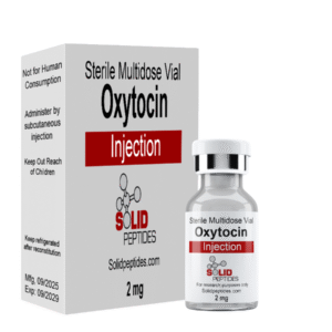
Hover to zoom
Identity Verified: LC-MS
(0 Reviews)
Oxytocin 10mg (6000 IU)
- Cyclic nonapeptide hormone with characteristic Cys1-Cys6 disulfide bridge, synthesized by Nobel laureate Vincent du Vigneaud (1954)
- OXTR agonist activating Gq/11-PLC-IP3/DAG cascade; modulates social cognition, stress axis, metabolism, and cardiovascular function
- Validated in ~25,000 publications spanning social neuroscience, reproductive physiology, psychiatry, metabolic medicine, and cardiology
- Mechanistic pathway studies
- In vitro receptor profiling
- HPLC verified identity and purity
$24.00In Stock
Ships same-day if ordered before 2PM EST
1
Encrypted Checkout
Global Express
Research Overview
Oxytocin has been extensively validated across multiple research domains:
- OXTR Signaling: Preferentially couples to Gαq/11 proteins, initiating canonical PLC-β cascade generating IP3 and DAG second messengers. Cryo-EM structural studies by Meyerowitz et al. (2022, Nature Structural & Molecular Biology) revealed magnesium coordination complex essential for receptor binding, with single cation-coordinating residue acting as molecular switch determining cation dependence
- PLC/IP3/DAG Cascade: IP3 triggers Ca²⁺ release from intracellular stores via IP3 receptors, amplified through store-operated Ca²⁺ entry (SOCE) and voltage-operated Ca²⁺ entry (VOCE) channels. DAG activates PKC, phosphorylating downstream targets. In uterine smooth muscle, calcium-calmodulin activates MLCK producing coordinated contractions. Additional calcium-independent mechanisms via RhoA/Rho kinase pathways contribute
- Downstream Signaling: Chatterjee et al. (2016) catalogued 66 molecules in oxytocin signaling network: 9 protein-protein interactions, 39 post-translational modifications, 14 protein translocation events, 22 activation/inhibition events. Key pathways include MAPK, CaMK, and PKC cascades converging on CREB and MEF-2 transcription factors
- Neuromodulatory Actions: Froemke & Young (2021, Annual Review of Neuroscience) established three-tiered model: (1) First-order: direct depolarization of principal neurons, (2) Second-order: activation of inhibitory interneurons reducing noise, (3) Third-order: regulation of dopamine/serotonin systems. Functions as attentional modulator increasing salience of social information rather than directly promoting prosocial behavior
- Social Behavior: Rigney et al. (2022) mapped neural circuits in paraventricular nucleus, supraoptic nuclei, bed nucleus of stria terminalis mediating maternal nurturing, social reward, pair bonding, social recognition. Facilitates long-term synaptic plasticity through experience-dependent neural reorganization
- Stress Axis Modulation: Althammer et al. (2021) documented inhibition of CRF mRNA expression at hypothalamus, reducing ACTH secretion and attenuating cortisol release. Parvocellular oxytocin neurons (~1% of oxytocin-producing neurons) function as "master cells" regulating entire oxytocin system
- Metabolic Effects: Ding et al. (2019, Obesity Reviews) established multi-pronged effects: promotes lipolysis, enhances beta-oxidation, reduces macrophage infiltration in adipose tissue, improves beta-cell responsivity, enhances insulin sensitivity. Long-acting selective OXTR agonists show antidiabetic and antiobesity effects
- Cardiovascular Protection: Jankowski et al. (2020) documented pleiotropic cardioprotective actions including ischemia-reperfusion injury protection via PI3K/Akt/RISK pathway, epicardial cell activation and heart regeneration, NLRP3 inflammasome inhibition, IL-1β/TNF-α suppression, nitric oxide release
- Bone Health: Breuil et al. (2021) reviewed anabolic effects on bone: promotes osteoblast differentiation via BMP-2 upregulation, dual effects on osteoclasts (promotes differentiation but inhibits resorptive capacity), prevents age-related sarcopenia
- Clinical Research: Meta-analysis by Zhang et al. (2025) of 12 RCTs with 498 ASD patients showed dose-dependent effects: at 48 IU/day, beneficial effects on social impairments emerged. Cai et al. (2018) meta-analysis of 5 RCTs with 223 participants found intranasal oxytocin "well tolerated and safe" with most common adverse events (nasal discomfort 14.3%, irritability 9.0%, tiredness 7.2%) not statistically different from placebo
Primary Research Applications
Social behavior neuroscience (pair bonding, maternal behavior, social recognition, oxytocin-dopamine/serotonin crosstalk)
GPCR signaling and receptor pharmacology (dual Gq/11 and Gi/Go coupling, biased agonism, cryo-EM structural studies)
Reproductive physiology (myometrial contraction, Ferguson reflex, milk ejection, pulsatile secretion patterns)
Psychiatric and neurodevelopmental disorders (ASD biomarker-defined subpopulations, schizophrenia negative symptoms, PTSD fear extinction)
Metabolic and obesity research (adipose tissue lipolysis, beta-cell function, glucose tolerance, long-acting OXTR agonist development)
Cardiovascular research (ischemia-reperfusion protection, epicardial cell activation, NLRP3 inflammasome inhibition)
Bone and musculoskeletal biology (osteoblast differentiation, BMD correlations, sarcopenia prevention)
Intranasal drug delivery (nose-to-brain transport pathway characterization, bioavailability optimization, neuroimaging studies)
Mechanism of Action
Oxytocin exerts its diverse physiological and behavioral effects primarily through the oxytocin receptor (OXTR), a Class A G protein-coupled receptor containing seven transmembrane domains.
1. OXTR and G Protein Coupling:
As comprehensively reviewed by Jurek & Neumann (2018, Physiological Reviews, ~25,000 publications catalogued), OXTR preferentially couples to Gαq/11 proteins, initiating canonical phospholipase C signaling. However, the receptor also couples to Gi/Go-type G proteins, enabling context-dependent signaling in different tissues. Meyerowitz et al. (2022, Nature Structural & Molecular Biology) cryo-EM structure revealed oxytocin binding disrupts transmembrane helix 7 (TM7), causing conformational changes facilitating G protein engagement. A single cation-coordinating residue acts as "molecular switch" determining magnesium-dependence.
2. Phospholipase C / IP3 / DAG Cascade:
Gq/11-mediated activation of phospholipase C-β (PLC-β) catalyzes hydrolysis of phosphatidylinositol 4,5-bisphosphate (PIP2) to generate two critical second messengers:
- Inositol 1,4,5-trisphosphate (IP3): Triggers Ca²⁺ release from intracellular stores via IP3 receptors on endoplasmic reticulum. Calcium entry further amplified through store-operated Ca²⁺ entry (SOCE) and voltage-operated Ca²⁺ entry (VOCE) channels. In uterine smooth muscle, calcium binds calmodulin and activates myosin light chain kinase (MLCK), producing coordinated contractions. Additional calcium-independent mechanisms via calcium sensitization through RhoA/Rho kinase pathways contribute
- Diacylglycerol (DAG): Remains membrane-associated and activates protein kinase C (PKC), which phosphorylates numerous downstream target proteins
3. Downstream MAPK, CaMK, and Transcriptional Regulation:
Chatterjee et al. (2016) systematically catalogued 66 molecules in oxytocin signaling network including 9 protein-protein interactions, 39 post-translational modifications, 14 protein translocation events, and 22 activation/inhibition events. Key pathways include MAPK (mitogen-activated protein kinase), CaMK (calcium/calmodulin-dependent kinase), and PKC cascades converging on transcription factors CREB (cAMP response element-binding protein) and MEF-2 (myocyte enhancer factor-2), regulating gene expression programs underlying long-term cellular adaptations including neurite outgrowth, cell viability, and survival.
4. Neuromodulatory Actions:
Froemke & Young (2021, Annual Review of Neuroscience) established oxytocin operates at three distinct levels in CNS:
- First-order modulation: Direct depolarization of principal neurons (e.g., hippocampal CA2 pyramidal cells)
- Second-order modulation: Activation of inhibitory interneurons to reduce noise and enhance signal-to-noise ratios in social information processing
- Third-order modulation: Regulation of other neuromodulatory systems including dopamine and serotonin
This multi-layered action facilitates long-term synaptic plasticity through experience-dependent neural reorganization, including disinhibition in auditory cortex during maternal behavior learning. Oxytocin acts as "attentional modulator, increasing salience of social information" rather than directly promoting prosocial behavior.
5. Stress Axis Modulation (HPA Axis):
Althammer et al. (2021) documented oxytocin inhibits corticotropin-releasing factor (CRF) mRNA expression at hypothalamus, resulting in reduced ACTH secretion and attenuated cortisol release during stress. Parvocellular oxytocin neurons (~1% of oxytocin-producing neurons) function as "master cells" regulating entire oxytocin system and mediating stress-buffering effects. This anxiolytic mechanism operates bidirectionally: stress activates oxytocin release, and oxytocin subsequently attenuates stress responses, creating homeostatic feedback loop.
“Mechanistic summaries on this page are provided for laboratory reference and should be interpreted within controlled experimental settings only.”
Preclinical Research Summary
Oxytocin has been validated in nearly 25,000 peer-reviewed publications spanning nine decades:
- Receptor Structural Biology: Meyerowitz et al. (2022) solved cryo-EM structure of OXTR-Gq signaling complex at high resolution, identifying magnesium coordination complex essential for oxytocin-receptor binding and discovering single amino acid position acting as molecular switch for cation dependence across vasopressin receptor family
- Social Behavior and Neural Circuits: Rigney et al. (2022, Endocrinology) mapped neural circuits in paraventricular nucleus, supraoptic nuclei, bed nucleus of stria terminalis mediating maternal nurturing, social reward, pair bonding, social recognition. Optogenetic and chemogenetic manipulation studies demonstrated oxytocin-dopamine crosstalk in ventral tegmental area and nucleus accumbens. Fiber photometry recording captured oxytocin release dynamics during social interactions
- Neuromodulation and Plasticity: Froemke & Young (2021) demonstrated oxytocin facilitates long-term synaptic plasticity through experience-dependent neural reorganization, including disinhibition in auditory cortex during maternal behavior learning in rodents. Established oxytocin as attentional modulator increasing salience of social information through three-tiered neuromodulatory action
- Stress Axis Modulation: Althammer et al. (2021) documented parvocellular oxytocin neurons function as "master cells" regulating entire oxytocin system. Oxytocin administration inhibits CRF mRNA expression at hypothalamus, reduces ACTH and cortisol in multiple stress paradigms (restraint, social defeat, predator exposure)
- Metabolic Effects: Ding et al. (2019) reviewed multi-pronged metabolic actions: promotes lipolysis and beta-oxidation in adipose tissue, reduces macrophage infiltration, improves beta-cell responsivity and glucose tolerance. Snider et al. (2019) demonstrated long-acting selective OXTR agonists show antidiabetic and antiobesity effects in male mice with dose-dependent reductions in body weight, improved glucose tolerance, and enhanced insulin sensitivity
- Cardiovascular Protection: Jankowski et al. (2020) documented ischemia-reperfusion injury protection through PI3K/Akt/RISK pathway activation in multiple cardiac injury models. Wasserman et al. (2022) showed oxytocin promotes epicardial cell activation and heart regeneration after cardiac injury. Demonstrated NLRP3 inflammasome inhibition, IL-1β and TNF-α suppression, nitric oxide release, and atrial natriuretic peptide-mediated protection
- Bone and Musculoskeletal: Breuil et al. (2021) reviewed anabolic effects on bone: promotes osteoblast differentiation via BMP-2 upregulation and estrogen-dependent pathways, dual effects on osteoclasts (promotes differentiation but inhibits resorptive capacity). Bone mineral density correlations with endogenous oxytocin levels in postmenopausal women. Prevents age-related sarcopenia in oxytocin knockout animal models
- Clinical Safety (Meta-analyses): MacDonald et al. (2011) reviewed 38 RCTs with 1,529 participants: intranasal oxytocin (18-40 IU) "produces no detectable subjective changes" and "produces no reliable side-effects." Cai et al. (2018) meta-analysis of 5 RCTs with 223 ASD participants found no statistically significant difference in adverse event rates between oxytocin and placebo. Most common: nasal discomfort (14.3%), irritability (9.0%), tiredness (7.2%)
- Clinical Efficacy (ASD): Zhang et al. (2025) meta-analysis of 12 RCTs with 498 ASD patients (252 oxytocin, 246 placebo): no significant overall effect at standard doses, but at 48 IU/day, beneficial effects on social impairments emerged. Dose-response analysis indicated higher doses more effective for both social impairments and repetitive behaviors. Sikich et al. (2021, NEJM, SOARS-B trial) 290 children/adolescents: no significant overall between-group differences, but subsequent biomarker analyses identified low baseline OXT subpopulations showing greater improvement
Pharmacokinetics: Quintana et al. (2021) reported intranasal bioavailability ~0.7-2%, with direct nose-to-brain transport accounting for >95% of brain-detected oxytocin. Plasma half-life 3-5 minutes for IV administration; intranasal brain concentrations peak at 30-60 minutes, return to baseline 90-120 minutes.
Academic References
- Jurek, B., & Neumann, I. D. (2018). The Oxytocin Receptor: From Intracellular Signaling to Behavior. Physiological Reviews, 98(3), 1805-1908. PMID: 29897293
- Meyerowitz, J. G., Rao, A., Sherrill, L., Snyder, J. C., Hwang, J. Y., Cinar, R., ... & Skiniotis, G. (2022). The oxytocin signaling complex reveals a molecular switch for cation dependence. Nature Structural & Molecular Biology, 29, 274-281. PMID: 35241813
- Froemke, R. C., & Young, L. J. (2021). Oxytocin, Neural Plasticity, and Social Behavior. Annual Review of Neuroscience, 44, 359-381. PMID: 33823654
- Rigney, N., de Vries, G. J., Petrulis, A., & Young, L. J. (2022). Oxytocin, Vasopressin, and Social Behavior: From Neural Circuits to Clinical Opportunities. Endocrinology, 163(8), bqac111. PMID: 35863332
- Ding, C., Leow, M. K-S., & Magkos, F. (2019). Oxytocin in metabolic homeostasis: implications for obesity and diabetes management. Obesity Reviews, 20(1), 22-40. PMID: 30253045
- Jankowski, M., Broderick, T. L., & Gutkowska, J. (2020). The Role of Oxytocin in Cardiovascular Protection. Frontiers in Psychology, 11, 2139. PMID: 32982875
This product is intended exclusively for in vitro laboratory research by qualified professionals. Not for human consumption. Not approved by the FDA.