Research Use Only: This product is supplied for laboratory research and in-vitro studies. Not for human or veterinary administration.
Hover to zoom
Identity Verified: LC-MS
(0 Reviews)
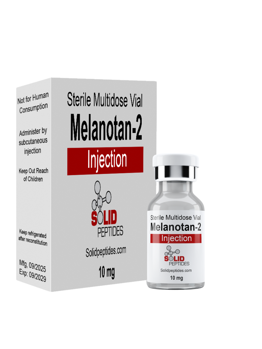
Melanotan-2 (10mg)
- Pan-Melanocortin Receptor Agonist: Cyclic heptapeptide with 100–1000× greater potency than native α-MSH at MC1R, MC3R, MC4R, and MC5R; ~33h half-life, ≥98% purity
- Melanogenesis & Photobiology Research: MC1R/cAMP/PKA/MITF cascade; UV-independent eumelanin biosynthesis and nucleotide excision repair pathway investigation
- MC4R Hypothalamic & Neuroprotection Research: Energy homeostasis circuits, adipose tissue lipolysis, peripheral nerve regeneration; validated in 400+ peer-reviewed publications
- Mechanistic pathway studies
- In vitro receptor profiling
- HPLC verified identity and purity
$40.00In Stock
Ships same-day if ordered before 2PM EST
1
Encrypted Checkout
Global Express
Research Overview
Melanotan-2 (MT-II) is a synthetic cyclic heptapeptide analog of alpha-melanocyte-stimulating hormone (α-MSH), developed at the University of Arizona in the early 1990s as an investigational compound for melanocortin receptor pharmacology research. Through lactam bridge cyclization between Asp5 and Lys10, D-Phe7 substitution, and norleucine replacement of Met4, the compound achieves a receptor potency 100–1000× greater than native α-MSH, near-complete resistance to proteolytic degradation, and an extended in vivo half-life of approximately 33 hours. Supplied as a lyophilized powder for in vitro research and intended strictly for qualified researchers, MT-II is a non-selective pan-agonist at all five melanocortin receptor subtypes (MC1R–MC5R) and has been validated in over 400 peer-reviewed publications spanning melanogenesis, energy homeostasis, neuroprotection, vascular biology, and alcohol use disorder neuropharmacology.
The compound's pharmacological versatility reflects the broad tissue distribution of melanocortin receptors. MC1R-mediated research has characterized the cAMP/PKA/CREB/MITF signaling cascade governing eumelanin biosynthesis, UV-independent melanogenesis, and nucleotide excision repair (NER) of UV-induced DNA damage. The moderate lipophilicity and cyclic structure of MT-II enable blood-brain barrier penetration, permitting MC4R activation in the hypothalamic paraventricular nucleus and lateral hypothalamus — a feature exploited in research on energy homeostasis circuits, adipose tissue lipolysis, and central-peripheral metabolic crosstalk. MC4R agonism also engages NF-κB suppression and anti-inflammatory signaling in glial and Schwann cells, providing neuroprotective effects demonstrated across sciatic nerve crush, cisplatin neuropathy, and brain ischemia models.
MT-II served as the structural template for two approved drugs: afamelanotide (Scenesse, EMA-approved for erythropoietic protoporphyria) and bremelanotide (PT-141, approved 2019). MT-II itself has never been approved by any regulatory agency for human therapeutic use and is classified as a prohibited substance under the WADA 2025 Prohibited List. Certificate of Analysis (CoA) provided with every lot.
Primary Research Applications
Melanocortin Receptor Pharmacology Research
Melanogenesis and Pigmentation Biology
MC4R Hypothalamic Energy Homeostasis Studies
Adipose Tissue Lipolysis Research
Neuroprotection and Nerve Regeneration Models
Atherosclerosis and Vascular Inflammation Research
Alcohol Use Disorder Neuropharmacology
Antidepressant Target Investigation
Mechanism of Action
MC1R-Mediated Melanogenesis and Photoprotection
MC1R / cAMP / PKA / MITF Cascade — MT-II binds MC1R on melanocytes with high affinity (Ki = 0.67 nM), activating the Gs-protein/adenylyl cyclase/cAMP signaling cascade. Elevated cAMP activates protein kinase A (PKA), which phosphorylates the transcription factor CREB, driving upregulation of microphthalmia-associated transcription factor (MITF). MITF in turn drives expression of melanogenic enzymes — tyrosinase, TRP-1, and TRP-2/DCT — catalyzing conversion of tyrosine to photoprotective eumelanin via DOPA and dopaquinone intermediates. The pathway preferentially produces eumelanin over phototoxic pheomelanin. MC1R activation also enhances nucleotide excision repair (NER) of UV-induced DNA damage and activates Nrf2-dependent antioxidant defense mechanisms through cAMP-dependent pathways (Mun et al., 2023).
MC4R-Mediated Hypothalamic and Metabolic Signaling
PVN / LH / Sympathetic Outflow — MT-II's cyclic structure and moderate lipophilicity (partition coefficient 2.82 at pH 7.35) enable blood-brain barrier penetration. MC4R is predominantly expressed in the hypothalamic paraventricular nucleus (PVN), lateral hypothalamus, brainstem, and spinal cord. MC4R activation increases sympathetic nervous system activity, upregulates uncoupling protein 1 (UCP1) in brown adipose tissue (thermogenesis), and enhances peripheral glucose disposal by upregulating GLUT4 mRNA expression in skeletal muscle ~3-fold (Heijboer et al., 2005). Peripheral MT-II reduces adipose tissue compartments through lipid mobilization and sympathetic activation of white adipose tissue — pair-fed control animals losing equivalent body mass retained significantly more adipose tissue than MT-II-treated animals, indicating direct lipolytic mechanisms beyond caloric restriction (Strader et al., 2007).
Neuroprotection via NF-κB Modulation
NF-κB / TNF-α / Schwann Cell Signaling — MT-II modulates NF-κB-mediated transcription in neural, glial, and endothelial cells, reducing pro-inflammatory agent production including TNF-α and IFN-γ after injury. This mechanism underlies neuroprotective effects demonstrated across sciatic nerve crush injury recovery (bell-shaped dose-response at 20 µg/kg per 48h), cisplatin-induced toxic neuropathy protection, brain ischemia neuroprotection with broad therapeutic time window, and neuronal rescue from excitotoxic insults. Cai & Hruby (2016) documented inhibition of TNF-α and IFN-γ-induced NF-κB activation in Schwann cells as a peripheral nerve regeneration mechanism.
Anti-Atherosclerotic Vascular Effects
Macrophage M2 Polarization / Endothelial NO Signaling — Rinne et al. (2014) demonstrated MT-II shifts resident plaque macrophages toward anti-inflammatory M2 phenotypes, reduces metabolic activity in atherosclerotic plaques (18F-FDG uptake), and enhances endothelium-dependent relaxation and nitric oxide-mediated vasodilation. These vascular benefits occurred independently of metabolic parameters or plasma cholesterol, confirming direct anti-inflammatory mechanisms. Chronic melanocortin-3/4 receptor activation has also been shown to increase blood pressure in preclinical models, indicating complex cardiovascular pharmacology (do Carmo et al., 2011).
Mast Cell Activation (Off-Target Effect)
MRGPRB2 / Histamine H1 Receptor — MT-II activates mast cells through both MRGPRB2-dependent and MRGPRB2-independent pathways (Jain et al., 2018), triggering histamine release that binds H1 receptors and produces dose-dependent hypothermia. This effect was abolished in mast cell-deficient mice and substantially reduced by H1 receptor blockade, explaining the flushing, nausea, and temperature changes observed as common off-target effects in MT-II research studies.
“Mechanistic summaries on this page are provided for laboratory reference and should be interpreted within controlled experimental settings only.”
Preclinical Research Summary
MT-II has been investigated across human pilot studies and multiple in vivo animal model systems. Early Phase I clinical investigations by Dorr et al. (1996) and Wessells et al. (1998) provided foundational pharmacological characterization in humans at the 0.01–0.03 mg/kg dose range, documenting melanotropic activity and common adverse effects including nausea, somnolence, and spontaneous physiological responses. MT-II itself has never progressed through IND-enabling toxicology programs and has never received regulatory approval for human therapeutic use.
In metabolic and adipose tissue research, Heijboer et al. (2005) demonstrated that intracerebroventricular MT-II (225 ng over 24 hours) significantly increased insulin-stimulated glucose disposal in hyperinsulinemic-euglycemic clamp studies (151±20 vs. 108±20 µmol/min/kg, p<0.01), with ~3-fold upregulation of GLUT4 mRNA in skeletal muscle. Strader et al. (2007) showed peripheral MT-II altered adipose tissue compartments in high-fat-fed mice; pair-fed controls retained more adipose tissue, indicating direct lipolytic mechanisms. Zhang et al. (2010) demonstrated that intermittent MT-II infusion reduced overall adiposity by approximately 80% relative to controls, with elevated ACC1 phosphorylation in fat tissue suggesting enhanced fatty acid oxidation.
In neuroprotection, Ter Laak et al. (2003) showed subcutaneous MT-II at 20 µg/kg per 48 hours significantly enhanced sensory function recovery after sciatic nerve crush injury in Wistar rats (bell-shaped dose-response) and partially protected against cisplatin-induced toxic neuropathy. In vascular research, Rinne et al. (2014) demonstrated 4-week MT-II infusion in LDLR-/-;ApoB100/100 mice reduced 18F-FDG uptake in atherosclerotic plaques, shifted macrophages toward M2 phenotypes, and enhanced endothelium-dependent relaxation independent of metabolic parameters or plasma cholesterol. In melanoma biology, Wu et al. (2020) showed topical MT-II reduced B16-F10 tumor volume to approximately 50% of control (989 vs. 2,017 mm³) via MC1R-dependent PTEN upregulation and AKT/NF-κB inhibition. Safety concerns include case reports of melanocytic lesion changes, at least five cutaneous melanoma cases (Bohm et al., 2025), rhabdomyolysis, and renal infarction in individuals who self-administered unregulated MT-II products. No comprehensive LD50, chronic toxicity, or genotoxicity studies have been published.
Academic References
- Dorr RT et al. (1996). Evaluation of melanotan-II, a superpotent cyclic melanotropic peptide in a pilot phase-I clinical study. Life Sciences.
- Wessells H et al. (1998). Synthetic melanotropic peptide initiates erections in men with psychogenic erectile dysfunction: double-blind, placebo controlled crossover study. Journal of Urology.
- Ter Laak MP et al. (2003). The potent melanocortin receptor agonist melanotan-II promotes peripheral nerve regeneration and has neuroprotective properties in the rat. European Journal of Pharmacology.
- Heijboer AC et al. (2005). Intracerebroventricular administration of melanotan II increases insulin sensitivity of glucose disposal in mice. Diabetologia.
- Strader AD et al. (2007). The effects of the melanocortin agonist (MT-II) on subcutaneous and visceral adipose tissue in rodents. Journal of Pharmacology and Experimental Therapeutics.
- Rinne P et al. (2014). Pharmacological activation of the melanocortin system limits plaque inflammation and ameliorates vascular dysfunction in atherosclerotic mice. Arteriosclerosis, Thrombosis, and Vascular Biology.
- Zhang Y et al. (2010). Intermittent MTII application evokes repeated anorexia and robust fat and weight loss. Peptides.
- Wu JC et al. (2020). Topical MTII Therapy Suppresses Melanoma Through PTEN Upregulation and Cyclooxygenase II Inhibition. International Journal of Molecular Sciences.
- Jain S et al. (2018). Melanotan II causes hypothermia in mice by activation of mast cells and stimulation of histamine 1 receptors. American Journal of Physiology - Endocrinology and Metabolism.
- Navarro M et al. (2015). Melanocortin Receptor Agonist Melanotan-II Synergistically Augments the Ability of Naltrexone to Blunt Binge-Like Ethanol Intake in Male C57BL/6J Mice. Alcoholism: Clinical & Experimental Research.
- Bohm M et al. (2025). An overview of benefits and risks of chronic melanocortin-1 receptor activation. Journal of the European Academy of Dermatology and Venereology.
- Markov DD et al. (2023). The Melanocortin System: A Promising Target for the Development of New Antidepressant Drugs. International Journal of Molecular Sciences.
This product is intended exclusively for in vitro laboratory research by qualified professionals. Not for human consumption. Not approved by the FDA.